You are here

Blogs

Which splice junction to target, predicting cryptic splice sites, and doing the two-non-overlapping oligo specificity experiment

I've had the pleasure of corresponding with Yuhong Liang (梁雨虹)of 4A Biotech in China; she is the product manager for Morpholino oligos at 4A Biotech. We have been working with 4A for a little over a year now and Yuhong has been learning very quickly, but is still new enough to the Morpholino field that she is asking questions of broad interest. She had some questions regarding which splice junction to target, predicting cryptic splice sites, and the process of doing the two-non-overlapping oligo specificity experiment. I am trying to help her understand Morpholino techniques so she can help users directly in China, so I responded at some length. I'll post my response to her (with her kind permission) in hopes other Morpholino users might glean a useful idea.

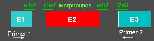
Yuhong was looking at a specific transcript with three exons, having a start in exon 1 and a stop in exon 3.

--------------------------------------------------------------

"As the most common outcome is exon skipping, we should consider i1e2 and e2i2 first, right?"

When targeting the first and last splice junctions, oligos usually cause intron inclusion. This sometimes provides a strong gene knockdown, so in some cases these will be very good choices (see the third figure and its discussion on this page: Splicing outcomes: targeting for exon skipping or intron inclusion). However, because removing exon 2 will frameshift downstream sequence it is also a very good choice; my favorite starting place is to skip the most upstream exon available that will cause a frameshift.

"What's the difference between i1e2 and e2i2?"

The i1e2 is a splice acceptor site and the e2i2 is a splice donor site. I generally prefer splice donor targets because there is some extra sequence conservation in the intron of the splice acceptor site which increases the chance of an off-target interaction. Acceptor sites have a polypyrimidine region and a nucleophilic A base that are involved in binding snRNPs and closing the splice lariat. All splice junctions have a few very conserved intronic bases; introns start with "gu" at the donor site and end with "ag" at the acceptor site. The additional polypyrimidine region and nucleophilic A base at the splice acceptor decrease the possible base sequences that an acceptor site can have, so there is some similarity between all of the acceptor sites in the transcriptome. That means the chance of an acceptor-targeted Morpholino binding to an unintended acceptor splice site is greater than the chance that a donor-targeted Morpholino will bind to an unintended donor splice site (because the donor sites can have more sequence variation). When someone asks for a Morpholino targeting a particular exon, I will try first to design a splice donor oligo and then if the oligo characteristics do not look good I will see if there is a better-looking splice acceptor. You can produce a stronger effect by using both donor and acceptor site oligos together at the same time; they should produce dose synergy.

"How can we confirm if there is cryptic splice site in exon2 before PCR?"

I don't know of a method for checking if there is a splice site prior to PCR. You can check the sequence to see whether there are AG or GT sites away from the splice junctions and these could be cryptic splice sites, but I think you will see many AG and GT sites which do not participate in splicing. Even the canonical GU starting an intron and AG ending the intron is not perfectly conserved; while the major spliceosome requires these bases, the minor spliceosome can use GU or AU at the acceptor and AG or AC at the donor site. There are some other semiconserved residues at active splice sites, for instance at a splice donor there is commonly an AG at the end of an exon just before the GU in the intron, but the exonic AG bases are not perfectly conserved -- their presence or absence give a hint about a cryptic site but not certainty. Cryptic splice acceptor sites need the polypyrimidine and nucleophilic A residues to function, but the configuration of these bases (e.g. distance from the junction) can be shifted about a bit.

The first method for checking whether or not a cryptic splice site is redirecting splicing is to do PCR and run the PCR products on an electrophoretic gel, comparing PCR products from Morpholino-treated and untreated cells/embryos/animals. If it is an easy splice, like successful clean excision of an exon, one gel can usually provide all the information needed but, if something unexpected occurs during splicing, then it might take several primer sets to determine what happened. A tricky situation is if a splice is redirected to a very close cryptic splice site. This can cause a very slight change in the migration of the PCR product band on the gel. This kind of short-distance splice-redirection can be revealed by sequencing the RT-PCR product; if a lab can afford to do the sequencing, it provides the most clear assessment of a splice oligo outcome. You see exactly where the cryptic splice site lies and you can predict if a frameshift occurred or not after oligo treatment.

"Are there two primers as below?"

That is the set that I would start with, and if it shows the expected outcome then that set is probably enough. If there is an unexpected intron inclusion, it might take a primer targeted in the intron to show that the inclusion has occurred.

"If designing for a wild splice outcome, how many Morpholino oligos should be needed?"

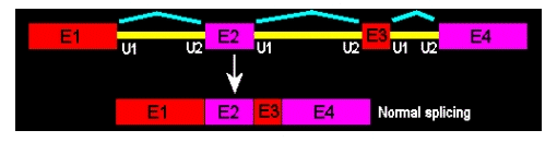
Wild-splice is the outcome when no Morpholino oligos are used. Think of "wild-type" vs. "mutant"; for splicing Morpholinos, we can talk of "wild-spliced" vs. "morphant" or “exon-skipped”. Here, no exon-skipping occurred; this is the mRNA as it is used by the cells to make functional proteins.

"About the two-nonoverlapping-oligos: should the oligos be used in separate experiments or by co-administration? When co-administrating, what concentration should be used?"

There are several possibilities for the non-overlapping oligo specificity control experiment.

First, try the oligos one-at-a-time, in separate experiments. If the morphants phenocopy each other, then that suggests that the oligos are specific and the observed phenotype is a result of interaction of the Morpholinos with their intended target; the probability is low that the same phenotype would be produced from both oligos due to interaction with an unintended RNA. However, there are RNA families that require additional concern; for instance, if a gene was duplicated and over many generations the RNA sequences from each duplicate began to diverge, but this happened recently enough that the sequence has not changed very much, then it is possible that two Morpholinos targeting a single RNA might both interact with the closely-related RNA produced by the gene duplication.

Next, you can try the two oligos together to see if there is dose-synergy. This is not done routinely, but is a good specificity experiment. Dose-synergy means that there is a greater-than-additive effect if the oligos are used together. If you get the same outcome using one of the oligos at some dose, let's call it one unit, then you get the same outcome if you use both oligos together each at 0.5 unit dose, then that does not show synergy, it is simply additive: 0.5U + 0.5U = 1U. However, if you use 0.25U of each oligo and you get the same outcome that you see from a single oligo at 1 U, that is an example of dose synergy: 0.25U + 0.25U < 1U (but caused the same outcome). Dose synergy is an indication that the oligos are interacting with the same mRNA (although it is still possible that they are interacting with two different RNAs in the same biochemical or physiological pathway). Dose synergy was proposed as a test for oligo specificity in this paper:
Bill BR, Petzold AM, Clark KJ, Schimmenti LA, Ekker SC. A primer for morpholino use in zebrafish. Zebrafish. 2009 Mar;6(1):69-77.
http://www.liebertonline.com/doi/pdfplus/10.1089/zeb.2008.0555

Base the initial doses for the co-administration dose-synergy experiment on a low dose of the single oligo that produces a phenotype. There is a factor that makes this more complicated, which is that different oligo sequences will have different effective dose ranges. Because of this, both oligos need to be tested individually to find their effective range, especially to determine the lower end of that effective range. Let's call those low effective doses U1 and U2 for oligo 1 and oligo 2. If you try a dose synergy experiment at 0.25 doses, you would use 0.25U1 of oligo 1 and 0.25 U2 of oligo 2. This corrects for the difference in activity of the two oligos.

You can also try the two oligos at 1 U of dose each, co-injected, to look for the effect of a stronger knockdown than is available with a single oligo.

There is a new sequence specificity experiment emerging, which uses a null-mutant embryo and a Morpholino targeting the RNA with the null-mutation; this has so far been explored in zebrafish embryos. If a Morpholino targeting a particular gene's RNA produces a measurable phenotype in a wild-type embryo, and then that Morpholino is injected into a mutant null for that gene and no change in phenotype is seen, this supports the hypothesis that the phenotype of the Morpholino in the wild-type is due to a specific interaction with the targeted RNA. If, however, the Morpholino still produces some phenotype in the null-mutant background, that does not preclude specificity; it is possible, for instance, that the Morpholino is interacting with maternally-deposited RNAs (this can be tested by injecting the Morpholino into embryos from a homozygous-null mother).

Sometimes a mutant does not produce the same phenotype as a wild-type embryo injected with a Morpholino targeting the gene that is null in the mutant. However, if that Morpholino sequence is injected into the null-mutant embryo then the Morpholino phenotype can disappear. That is the case if the mutant is compensating for the loss of the gene of interest by altering the expression of other genes; this is homeostasis, the tendency of a stressed organism to readjust its physiology to attain a healthy state. When that Morpholino is injected into the null mutant, it has no functional target (the target is the null) and other genes have adjusted their expression to compensate for the loss of the null gene, so you don't see the Morpholino phenotype. This was initially explored in this paper:

Rossi A, Kontarakis Z, Gerri C, Nolte H, Hölper S, Krüger M, Stainier DYR. Genetic compensation induced by deleterious mutations but not gene knockdowns. Nature. 2015 Aug 13;524(7564):230-3. doi: 10.1038/nature14580. Epub 2015 Jul 13.
http://www.nature.com/nature/journal/vaop/ncurrent/full/nature14580.html

I recently had a nice discussion about this oligo specificity control strategy with Martin Blum, which is on my blog here:
Validating Morpholino phenotypes with CRISPRs

Getting Morpholinos into cultured cells

There are three usual options to consider for cell culture experiments with Morpholinos. There are other methods, but most labs choose one of these.

(1) If your cells can tolerate electroporation, that is an inexpensive option for delivery of standard Morpholino oligos. However, electroporation causes high cell mortality for many cell types.

(2) Use Endo-Porter to deliver a standard Morpholino oligo. Endo-Porter is most effective in its DMSO formulation. The Endo-Porter peptide is poorly soluble in water and when the DMSO formulation is pipetted into culture medium, it is important to immediately swirl the medium to disperse the Endo-Porter. The Endo-Porter peptide will form complexes in the aqueous medium and swirling keeps these complexes small and abundant; these small and abundant complexes produce the highest delivery efficacy. The Endo-Porter aqueous formulation is provided because some undifferentiated cell types are triggered to differentiate by small concentrations of DMSO. The aqueous formulation is about a quarter as effective as the DMSO formulation and should only be used if needed. To treat around 150 ml of culture medium, you will need one order of Endo-Porter ($200) and 300 nanomoles of standard Morpholino ($400). The 150 ml estimate can vary, as different cell lines have different optimal concentrations of Endo-Porter and the required dose of a Morpholino oligo will vary with the sequence; standard Morpholinos are typically added to a final concentration of 2 to 10 micromolar in the cell medium.

(3) Vivo-Morpholinos can be used in cell cultures by adding the Vivo-Morpholino to the medium on the cells, with no other delivery system required. Vivo-Morpholinos are typically added to a final concentration of 2 to 10 micromolar in the cell medium. Vivo-Morpholinos work with most cell types tested but some are sensitive to the Vivo-Morpholino's attached delivery moiety, an octaguanidinium dendrimer. 400 nanomoles of a Vivo-Morpholino costs $700, more than the combination of a standard Morpholino with Endo-Porter but you receive more oligo.

Validating Morpholino phenotypes with CRISPRs

I've been enjoying a conversation with Martin Blum about Morpholinos, CRISPR mutants, funding and publication. He wrote:

Good morning Jon,

My study section at the German Research Council DFG now accepts MO-proposals, no problem. Still, many people particularly working in zebrafish are too careless with controls and rescues. That might backfire at one point in the future.

My take is that we will use CRISPR to validate MOs and then use MOs most of the time.

Best wishes,
Martin

-----------------------------------------------------
My response
-----------------------------------------------------

Hi Martin,

You want a CRISPR mutant to validate the Morphlino, but we know compensation in the mutant can conceal the effect of losing the protein, which can be revealed by a knockdown. What do you do if the Morpholino shows a phenotype and the CRISPR doesn't? Is this validation useful? More below...

I heartily agree that specificity controls need to be tight. Five-mispair oligos are worthless. Rescues are very good if they work, but ectopic expression can mess them up. The two-nonoverlapping-oligo phenocopy is good but there have been cases where both oligos have off-target interactions, so that can take more oligos to nail down. Checking for dose synergy with two non-overlapping oligos is stronger when combined with the one-oligo-at-a-time phenocopy previously mentioned. But my favorite specificity control today is:

You have a Morpholino and it produces a phenotype when injected into a wild-type creature. You also have the CRISPR mutant for the same target gene, it has no phenotype. You inject the Morpholino into the mutant, you see no phenotype. That tells us that compensation in the mutant is concealing the phenotype that the Morpholino reveals, and further tells us that the Morpholino phenotype is due to interaction with the targeted transcript and not an unexpected RNA. If that is the validation of a Morpholino with a CRISPR mutant that you are proposing, then I am enthusiastically on-board.

Alex reviewed this message prior to sending and pointed out that if the Morpholino has changed physical state, e.g. through aggregation in the solution state, then relying on a negative result (no Morpholino phenotype in the mutant) could be dangerous. To avoid that, the oligo should be injected to wild-type embryos and mutant embryos in the same session, so if there is an issue with the oligo's physical state then you won't see the Morpholino phenotype in the wild-type embryo.

There is a confounding problem that could show up for early phenotypes. If a heterozygous mother is bred to produce a homozygous mutant embryo, there could be some wild-type maternal transcript that a translation-blocking Morpholino could shut down. This might lead to a more extreme early phenotype in the Morpholino-injected embryos than in the homozygous mutants from heterozygous mothers, which have some wild-type maternal transcript. In this case, the Morpholino could be specific but the phenotype would persist in the mutant background.

I'll post this discussion in my blog; if you like I would be happy to credit you with stimulating this response (I won't do so unless you tell me to -- I could simply add to the blog your message in order to set up the response).

Best wishes to you too,

- Jon

-------------------------------------------------

Hi Jon,

Thank you so much for this reasoning on controls, and please: post it on you blog. I am aware of all you are saying, and we do struggle with specificity controls in some cases quite a bit. So far we have always managed to prove it and to convince referees of manuscripts.

With best wishes,
Martin

-------------------------------------------------

Note (Jon): I first became aware of a Morpholino-in-a-mutant strategy from Didier Stainier's group's paper on genetic compensation in morphants:
Rossi A, Kontarakis Z, Gerri C, Nolte H, Hölper S, Krüger M, Stainier DYR. Genetic compensation induced by deleterious mutations but not gene knockdowns. Nature. 2015;[Epub ahead of print] doi:10.1038/nature14580
http://www.nature.com/nature/journal/vaop/ncurrent/full/nature14580.html

Assessing activity of a splice-modifying Morpholino

This is about detecting the activity of a splice-modifying oligo by reverse-transcriptase PCR and gel electrophoresis.

The expected outcome of an exon targeting an exon 3 splice junction (either i2e3 or e3i3) is elimination of exon 3 from the mature mRNA (along with both of its flanking introns, i2 and i3). The typical way to assess activity of a splice-modifying oligo is to isolate RNA from treated samples and run reverse-transcriptase PCR (RT-PCR), then run the PCR product on a gel to assess the mass of the product against a DNA ladder.

I have some notes on the expected outcomes of splice-modifying oligos (and a description of some possible unexpected outcomes) elsewhere on my blog:

Splicing outcomes: targeting for exon skipping or intron inclusion

To detect elimination of exon 3, we would typically design primers to exons 2 and 4, set back from the e2i2 and i3e4 junctions sufficiently that the RT-PCR product without exon 3 will still be about 100 bases long (that way enough dye binds in the RT-PCR product to see it clearly on a gel; that's tough if the fragment is too short). For short exons, it might be necessary to place the primers more distantly, for instance in exons 2 and 5. If the product is subject to nonsense-mediated decay, you might not see the splice-modified band; instead, NMD is visualized as a dimming or disappearance of the band when compared to the wild-spliced (negative control) band. To compare band intensities, load the wells lightly so the bands are not saturated and compare with RT-PCR of a housekeeping gene to confirm your total RNA loading is comparable between wells. If the wells are overloaded (so the bands are saturated), you might not see a partial knockdown of the wild-spliced band.

(review) Loss-of-function genetic tools for animal models: cross-species and cross-platform differences

Housden BE, Muhar M, Gemberling M, Gersbach CA, Stainier DY, Seydoux G, Mohr SE, Zuber J, Perrimon N. Loss-of-function genetic tools for animal models: cross-species and cross-platform differences. Nat Rev Genet. 2016 Oct 31. doi: 10.1038/nrg.2016.118. [Epub ahead of print]

Our understanding of the genetic mechanisms that underlie biological processes has relied extensively on loss-of-function (LOF) analyses. LOF methods target DNA, RNA or protein to reduce or to ablate gene function. By analysing the phenotypes that are caused by these perturbations the wild-type function of genes can be elucidated. Although all LOF methods reduce gene activity, the choice of approach (for example, mutagenesis, CRISPR-based gene editing, RNA interference, morpholinos or pharmacological inhibition) can have a major effect on phenotypic outcomes. Interpretation of the LOF phenotype must take into account the biological process that is targeted by each method. The practicality and efficiency of LOF methods also vary considerably between model systems. We describe parameters for choosing the optimal combination of method and system, and for interpreting phenotypes within the constraints of each method.

http://www.nature.com/nrg/journal/vaop/ncurrent/full/nrg.2016.118.html

Acute RNAi versus knockout: mutation triggers compensation

No Morpholino work in this one, but it explores a difference between targeting RNA versus DNA.

Cell-Intrinsic Adaptation Arising from Chronic Ablation of a Key Rho GTPase Regulator.
Cerikan B, Shaheen R, Colo GP, Gläßer C, Hata S, Knobeloch KP, Alkuraya FS, Fässler R, Schiebel E.
Dev Cell. 2016 Sep 28. pii: S1534-5807(16)30595-0. doi: 10.1016/j.devcel.2016.08.020.

http://www.sciencedirect.com/science/article/pii/S1534580716305950

"Thus, phenotypes of gene inactivation are critically dependent on the timescale, as acute knockdown reflects a transient state of adjustment to a new equilibrium that is attained following compensation."

Morpholino drug approved by FDA

The US Food and Drug Administration today (19 Sep 2016) granted Accelerated Approval to eteplirsen (EXONDYS 51), a Morpholino oligo-based treatment for some forms of Duchenne muscular dystrophy (DMD). This is the first approval of a Morpholino drug.

DMD, a devastating childhood disease that is usually fatal within the first 20 years of life, is caused when the protein dystrophin is not produced. Eteplirsen is a Morpholino antisense oligo targeting exon 51 of the DMD transcript to cause its excision from the DMD pre-mRNA. Some mutations causing DMD are frameshift mutation (often deletions) occurring adjacent to exon 51; for some of these, skipping exon 51 can restore the reading frame of the functional dystrophin protein and cause some internally-truncated dystrophin to be made.

http://www.fda.gov/NewsEvents/Newsroom/PressAnnouncements/ucm521263.htm
http://investorrelations.sarepta.com/phoenix.zhtml?c=64231&p=irol-newsAr...
http://www.bloomberg.com/news/articles/2016-09-19/sarepta-wins-approval-...

Eteplirsen (sequence source: US FDA ETEPLIRSEN BRIEFING DOCUMENT NDA 206488)

Morpholino phosphorodiamidate antisense oligomer

CTCCAACATCAAGGAAGATGGCATTTCTAG
20-mer
20% G
43% CG
Predicted Tm: 88.9°C at 10 µM oligo.

Oligo complement
CTAGAAATGCCATCTTCCTTGATGTTGGAG

DMD-001 Exon 51, ENST00000357033.8 in Ensembl.org, RNA target site marked. Given that the target site is within an exon, this is likely blocking binding of an exonic splice enhancer protein and so altering splicing by interfering with splice regulation.
CTCCTACTCAGACTGTTACTCTGGTGACACAACCTGTGGTTACTAAGGAAACTGCCATCT
CCAAA[CTAGAAATGCCATCTTCCTTGATGTTGGAG]GTACCTGCTCTGGCAGATTTCAACC
GGGCTTGGACAGAACTTACCGACTGGCTTTCTCTGCTTGATCAAGTTATAAAATCACAGA
GGGTGATGGTGGGTGACCTTGAGGATATCAACGAGATGATCATCAAGCAGAAG

miRNA targeting and second oligo specificity controls

Once again, a conversation with a new Morpholino user led to a discussion others might find useful.

For the two non-overlapping oligo specificity control, you do two separate sets of injections of the two oligos targeting the same miRNA. If the embryos treated with the oligo targeting the 5' end of the miRNA produces the same phenotype as the oligo targeting the 3' end of the miRNA, then that is good: it supports the idea that the phenotype you are seeing is caused by knocking down the activity of the miRNA you intend to target, and not caused by binding to an unexpected RNA.

You might think that only the oligo that targets the guide strand of the miRNA would give a phenotype. The reason both of the oligos should work is that Morpholinos can invade the pre-miRNA and pri-miRNA before they have been processed into the mature double-stranded miRNA. Either one of the oligos can invade the immature miRNA hairpin and once the oligo is bound there is no longer a double-stranded RNA to be processed and become the mature miRNA. By opening up the hairpin and displacing part of the RNA, the Morpholino is acting as an inhibitor of miRNA processing enzymes (e.g. Drosha, Dicer). If both oligos can inhibit maturation of the miRNA, loss of the mature miRNA should produce the same phenotype in the embryos.

Kloosterman WP, Lagendijk AK, Ketting RF, Moulton JD, Plasterk RHA. Targeted inhibition of miRNA maturation with morpholinos reveals a role for miR-375 in pancreatic islet development. PLoS Biol. 2007;5(8): e203.
http://journals.plos.org/plosbiology/article?id=10.1371/journal.pbio.005...

Pages

Subscribe to RSS - blogs